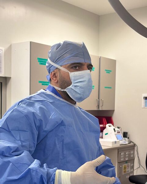
Angioplasty and stents
Coronary angioplasty widens a narrowed heart artery from the inside using a small balloon, and a stent, a fine metal mesh tube, helps keep it open.
Read the guideIntravascular ultrasound (IVUS) uses a tiny ultrasound probe on a catheter to image a heart artery from the inside. It shows the size of the artery, the plaque in its wall and how well a stent has expanded. Dr Shady Habboush uses IVUS during angioplasty at Saudi German Hospital Dubai when it can help guide treatment.
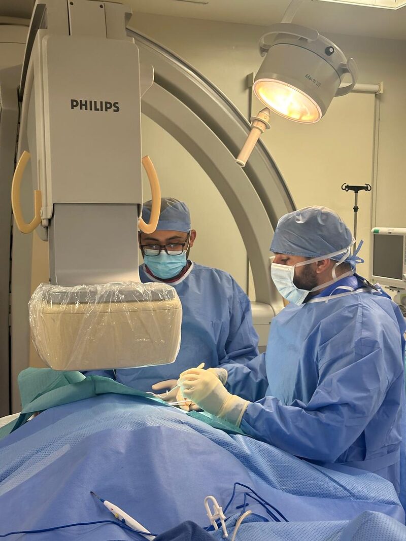
IVUS adds a small extra risk to the procedure, mainly a spasm or, rarely, an injury to the artery from the catheter. It is not needed for every stent. Whether imaging is used is decided case by case, and you can ask about it before the procedure.
IVUS is done during an angiogram or angioplasty, through the same catheter route.
A thin imaging catheter is passed along a guide wire into the artery.
The probe is pulled back slowly, recording cross-sectional images of the artery wall.
The images are used to choose the stent size and to check the result after the stent is placed.
It usually adds only a few minutes to the procedure.
You will not notice IVUS itself: recovery is the same as for the angiogram or angioplasty.
The 2024 European guidelines for chronic coronary syndromes recommend intravascular imaging, such as IVUS or OCT, to guide stenting in complex narrowings, for example the left main artery, branch points and long narrowings.
No. It is part of an angiogram or angioplasty and uses the same access route.
IVUS itself uses ultrasound, not X-ray, and does not need extra contrast dye. The procedure around it still uses X-ray.
No. It is mainly used for complex or unclear narrowings, as the guidelines recommend. Dr Shady will tell you if it is planned.
Both image the artery from inside. IVUS uses sound waves and sees deeper into the wall; OCT uses light and gives very fine detail near the surface.
Coronary angioplasty widens a narrowed heart artery from the inside using a small balloon, and a stent, a fine metal mesh tube, helps keep it open.
Read the guideA coronary angiogram is an X-ray test that shows the arteries that supply the heart.
Read the guideCoronary artery disease is narrowing of the heart's arteries by fatty plaque.
Read the guideMedical facts on this page follow the sources below.
Medically reviewed against ESC and NHS guidance · Updated October 2026. General information, not medical advice.
Appointments are made through Saudi German Hospital Dubai, Hessa Street, Al Barsha 3. Consultations in Arabic, English and German.
Chest pain right now? Do not wait for an appointment. Call 998 or go to the nearest emergency department.